|
FAQs
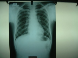
Plain X-Rays & Digital X-Rays Films
What are X-rays?
X-rays are electro-magnetic radiation,
which are produced by special machines called X-ray machines. These cannot be
seen, felt or heard.
 How do X-rays work? How do X-rays work?
Different parts of the body behave differently with X-rays. Structures
such as bone absorb X-rays, whereas air in the lungs lets all X-rays
pass through. Thus, when X-rays pass through the body, when they come
out, they have different strengths, depending on what parts of the body
they have passed through. When these X-rays hit a film (like a
photographic film), that film gets exposed depending upon this
variation. Like a photographic film, this special film also needs to be
developed, before we can see the final picture.
Where are X-rays useful?
X-rays have been used to look at all parts of the body.
Specifically, they are required for the chest, all bones and joints and
for the abdomen.
Are there any dangers?
Since, X-rays involve radiation, there is a theoretical risk, though
none in practice. In women who are pregnant, X-rays should be performed
only after weighing all risks and benefits.
What are the dyes used with X-rays?
Sometimes, artifical dyes are used to improve our ability to see
internal structures. These usually form part of a "procedure".
The common dyes used are either barium containing (barium sulphate) or
iodine containing. Barium sulphate is used for all barium examinations
to study the stomach and intestines. Iodine containing dyes are usually
injected in the veins to study the kidneys, during angiography, etc.
Are there any complications of the dye?
5% of patients may get nausea and redness of skin. Though severe
reactions are known, these are very rare and uncommon. However, in
patients show have a previous history of allergy, those who are
asthmatics, those wich renal and cardiac failure, a special dye which is
more expensive, but safer should be used, to prevent a reaction.
Who is qualified to report X-rays?
Only radiologists are trained to read X-rays and all X-rays should carry
a radiologist's report. Other physicians and non-radiology centres may
also perform X-rays, but they are usually not qualified. Before going
for an X-ray, ask the centre, whether it will be done under the
radiologist's supervision.
Are there any newer advances in X-rays?
X-rays are used in CT scanning (computed tomography). Digital
radiography uses X-rays for directly producing images on a computer,
bypassing the film - this is very helpful in emergency situations,
such as the trauma centre or intensive care unit.

What are Digital X-rays?
All images obtained using digital systems are digital x-rays. The
commonest digital method used is the CR system that produces digital
X-rays on a computer. See the accompanying picture for an idea of how
this works.
What are the advantages of Digital X-rays?*
Digital X-rays are superior to conventional X-rays in resolution and
quality.
How much do X-rays cost?
A single X-ray costs Rs. 120. Procedures cost more as more X-rays are
taken.
*Digital X-Ray facility
starting shortly.
Ultrasound
 What is ultrasound? What is ultrasound?
Ultrasound is a technique which uses sound waves of high frequency to produce
images.
How does ultrasound work?
Different parts in the body have a different response to high-frequency
ultrasonic waves passed through them. When ultrasound waves are passed through
the body, many tissues reflect sound waves partially and transmit the rest,
which are then reflected back from deeper structures. The reflected waves are
measured and depending on the time it takes for them to return, the depth of the
echo is decided - the intensity decides the grayness of the area
Where is ultrasound useful?
Ultrasound is used in many parts of the body, specifically to look at the fetus,
for other gynecological abnormalities and to look at the abdomen, orbits,
thyroid gland, breast, testes, etc.
Are there any dangers?
There is no known danger to the use of ultrasound
Are there any dyes in ultrasound, as in X-rays?
As yet there are no "dyes", injected in routine ultrasound practice.
Contrast media however are being evaluated for use in clinical trials.
How
much does ultrasound cost?
Ultrasound examinations cost between Rs 300 and 600, depending on the area of
the body examined
Who is qualified to do ultrasound?
Only radiologists trained in ultrasound should perform these investigations. In
many situations, however, gynecologists also perform ultrasound examinations on
their own patients - if adequately trained, this is acceptable.
Are there any newer advances in ultrasound?
Newer advances include the following
- Higher resolution scanning for small areas such as the superfical
ligaments and tendons of the body
- Doppler - this allows examination of blood vessels
- 3D Ultrasound - this allow three dimensional studies of various parts of
the body
Color Doppler
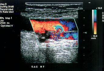 What
is Color Doppler? What
is Color Doppler?
Color Doppler is a special ultrasound technique, which allows us to evaluate
blood vessels.
What is the principle?
Using the Doppler principle of changing pitch with velocity, ultrasound waves
that reflect from the red blood corpuscles in arteries and veins are evaluated
for velocity and amplitude and color maps of the vessels can be generated.
Is special equipment required?
An ultrasound machine equipped with color Doppler facilities is required. These
are now readily available at many centers.
What is its utility?
Color Doppler is very useful in evaluating the carotid arteries in the neck, the
heart (echocardiography), the arteries and veins in the abdomen and the arteries
and veins in the upper and lower limbs.
What is power Doppler?
It is a type of color Doppler.
Is any special preparation
required?
No
What is the cost?
Between Rs 1000 and 2000.
Who is qualified to perform
color Doppler?
A qualified radiologist is the only one who should perform color Doppler
examinations of the vessels in the body.
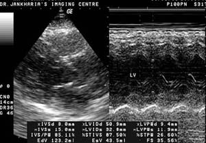
Echocardiography
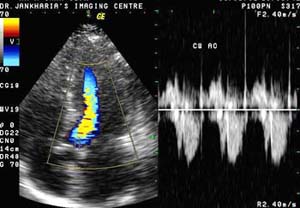 What is echocardiography?
It is a method of studying the heart and the adjacent great vessels
using ultrasound.
What is it used for?
Echocardiography is used to study the structure and function of the
chambers of the heart, the integrity of the valves and the coverings of
the heart.
Is any preparation required?
None.
Are there any dangers of echocardiography?
None.
How much does it cost?
Anywhere between Rs 600 and Rs 1200.
Does color Doppler help?
It allows better visualization of the vessels and the chambers of the
heart
Who is qualified to perform echocardiograms?
Trained cardiologists and radiologists.
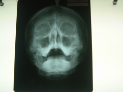
OPG & Cephalogram
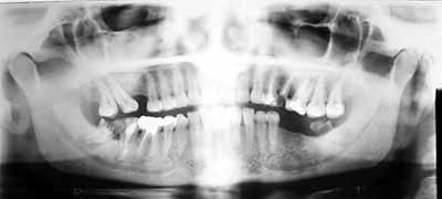 What
is OPG? What
is OPG?
OPG stands for Orthopantomography. It is a special method for obtaining
radiographs of the teeth-bearing jaws, both upper and lower.
How is it different from regular X-ray machines?
A regular X-ray machine cannot take detailed pictures of the jaw-bones. An OPG
machine is specially constructed so that it rotates around the jaw-bones, thus
giving us an extremely good idea about the structure of the jaw bones. Yes,
x-rays are used, but the method is totally different.
In what situations are they needed?
OPG x-rays are usually asked for by dentists, whether they be general dentists,
orthodontists, oral surgeons or prosthodontists/implantologists. Because OPGs
give a bird-eye view of the teeth and the adjacent bones, they are useful in a
wide-variety of conditions including infections, tumors, congenital
abnormalities, pre-implant evaluation and trauma.
Are they any risks?
Just as with x-rays elsewhere in the body, if a lady thinks she might be
pregnant, an OPG can be avoided. No other risks exist.
Is any dye injected?
No
How much time does it take to get OPGs done?
Around 10 minutes.
How much does it cost?
On an average, Rs 250.
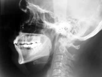 What
is a cephalogram? What
is a cephalogram?
It is an x-ray of the face, obtained so that accurate measurements of the face
can be performed.
What is its use?
Oral surgeons, orthodontists and prosthodontists need cephalograms prior to
planning surgeries and teeth manipulation.
How much time does it take?
On an average, 20 minutes.
How much does it cost?
Rs 300
Barium Studies
 What are barium studies? What are barium studies?
These are studies of the gastro-intestinal performed using barium
sulphate and x-rays.
Why are there different types of barium studies?
Depending on the area being examined, we have barium swallow, meal,
meal-follow-through, enema and small bowel enema.
What are these?
Barium swallow is a study for the esophagus, barium meal for the
stomach, barium meal follow-through for the small bowel, barium enema
for the large bowel and small bowel enema for the small bowel. In
swallow, meal and meal-follow-through examinations, the patient has to
drink barium. In barium enema examinations, barium is injected using an
enema tube. In small bowel enema examinations (enteroclysis), a tube is
inserted from the nose to the duodenum and barium is injected.
How do barium examinations work?
Barium is an inert substance that coats the internal lining of the bowel
and fills up its lumen. It is radio-opaque and thus seen very well on
x-rays.
Is there any danger?
Barium by itself is an inert substance and completely harmless. However
if it escapes into the abdominal or thoracic cavity through a
perforation, it can cause severe inflammation. Thus barium studies
should not be done in patients with suspected perforation.
What preparation is required?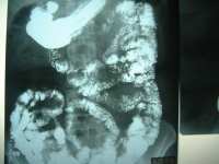
For barium swallow, none. For barium meal, at least six hours fasting.
For barium meal follow-through, overnight fasting with Dulcolax tablets
for clearing the bowel. For small bowel and barium enema, overnight
fasting with liquid diet the day before and aggressive clearing of the
bowel with Dulcolax tablets the night before and in the morning.
How much time do they take?
From 15 minutes for a swallow to 2-3hours for barium meal follow-through
examinations.
How much do these studies cost?
Between Rs 800 and Rs 2500.
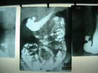
Barium Study - Enema Barium Study - Bowel Enema
Other Radiology Procedures
IVU
(intravenous urography)
In this, a dye is injected intravenously and x-ray pictures of the kidneys,
ureters and bladder are obtained. The dye is radio-opaque and seen well with
x-rays. Overnight fasting and good preparation of the colon with Dulcolax are
required. The cost is between Rs 1000 to 2500.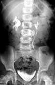
MCU (micturating cystourethrography)
Dye is introduced into the urinary bladder and the patient is asked to micturate/urinate.
X-ray pictures are obtained during the act of micturition to assess the function
and structure of the urinary bladder and urethra.
RGU (retrograde urethrography)
Dye is injected through the urethra from the glans penis and x-ray pictures are
taken. This helps in assessing the urethra and the bladder base.
Fistulogram and sinusogram
In these studies, using a small catheter, iodinated dye is injected into the
cutaneous sinus or fistula and x-rays are taken, which help in identifying the
tract of the sinus or fistula.
Sialography
In this, the parotid duct is cannulated from the mouth and x-ray pictures of the
parotid duct and gland are obtained.
Angiography, venography
The arteries are catheterized usually through the femoral artery and after
injection of iodinated dye, x-rays are taken. If the same study is performed for
the veins, we get venograms.
HSG (hysterosalpingography)
The cervix is cannulated and iodinated dye is injected into the cervical and
uterine lumen. The Fallopian tubes are then well seen. This procedure is used to
study the patency of the passage as well as other structural abnormalities.
|